
WallsHeaven makes decorative wall murals in high quality printing

32,956 Photomicrograph Wall Murals - Canvas Prints - Stickers

pine wood micrograph
pine cone and pine ovulate on the slide under microscope view.
amoeba proteus / chaos diffluens
realistic computer-generated micrograph showing a group of coronavirus particles as seen under a tra
microscopic view of lung tissue with congestion as shown by the red blood cells in the walls of the
microscopic view of mesothelioma cancer cells infiltrating the beige fibrous lung pleura.
nanoparticles made of lipids (liposomes) containing an anticancer drug (doxorubicin)
spinal cord cross section under the microscope showing neurones and glial cells (nervous tissue) - o
prostate cancer(turp): microscopic image of prostatic tissue, adenocarcinoma, malignant neoplasm,aty
rapeseed flower bud halve, cross section, 20x light micrograph. brassica napus subsp. napus, also kn
capillary hemangioma, light micrograph
lung adenocarcinoma, light micrograph
camera photo of endometrioid carcinoma with squamous differentiation, magnification 200x, photograph
skin tissue biopsy: lichen planus. show skin tissue, epidermis show hyperkeratosis, acanthosis, saw-
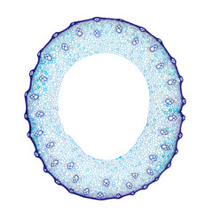
wheat stem, cross section, 8x light micrograph. blue stained stem of triticum under a light microsco
pine leaf, cross section, 20x light micrograph. pine tree needle under the microscope. epidermis cel
medical themes: transbronchial lung byopsia of cryptogenic organizing pneumonia formerly known as br
liver with macrovesicular steatosis displaying hepatocytes containing large fat droplets. stained us
peripheral nerve histology: bundles of myelinated axon fibers.
photomicrograph of lung tissue with silicosis pathology under a microscope, revealing silica particl
nanotechnology, medicine, nanoparticles, targeted therapy, drug delivery, healthcare, biomedical, in
cervical lymph node(biopsy): non-hodgkin's lymphoma-high grade. light microscopically show monotonou
tendon sheath (biopsy): chronic synovitis. smear show area of fibrosis and infiltration of inflammat
backgrounds of human cells tissue of lung human under the microscope in pathology lab.view in micros
a closeup captured the complex dance of mitochondria within a cell, their energy production a tiny b
polarization microscopy of copper sulfate crystals at 100x.
microscope image of spider silk thread fibers
backgrounds of human cells tissue of cervix under the microscope in pathology lab.view in microscopi
d of escherichia coli bacteria colony in a petri dish
backgrounds of human cells tissue of cervix under the microscope in pathology lab.view in microscopi
a scanning electron micrograph of the head of a cricket.
this is a photomicrograph of corn seeds.this photo focuses on the germ sheath cells of corn.magnify
endocrine pancreas. islets of langerhans
urinary bladder. transitional epithelium
human fat body tissue under microscope view for physiology education
a captivating microscopic view of a single strand of hair, revealing its structure, texture, and uni
glowing fibers of the hairy calyptra of polytrichum moss.
cell wall thickenings in a branch leaf of sphagnum squarrosum.
glowing cell walls of a flattened moss operculum, with polarization.
plant micrograph background
abstract micrograph of chopped moss leaves, with polarization.
starch granules of common ginger (zingiber officinale) tuber. stained by lugol iodine. objective 40x
neural network artistic representation
close-up of colorful concentric patterns on a reishi mushroom's surface exhibiting natural textures
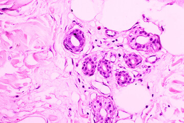
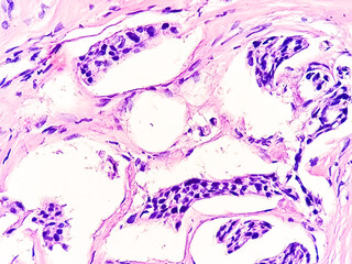
breast biopsy photomicrograph of infiltrating duct cell carcinoma (nos), grade-1
plant cell micrograph, microscopic view of cells enlarged
close-up of a detailed green leaf with visible veins and texture, isolated on a white background.
a single cancer cell on a blue surface
hereditary hemolytic anemia hemoglobin e hb e disease with hypochromia anisopoikilocytosis pencil ce
lymph node. hodgkin lymphoma
toxic goiter light micrograph , thyroid gland, enlarged, hyperthyroidism, pathology
tilia stem, cross section, 8x light micrograph. stem of basswood, also known as linden, under the li
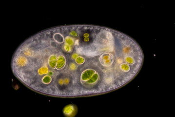
study of protozoa and algae under the microscope for education.
a 50x magnification photomicrograph of paramecium caudatum's animal conjugation
vibrant purple medical marijuana flower with intricate orange hairs and surrounding leaves, showcasi
splenic capsule hyaline degeneration, light micrograph, photo under microscope
a close-up microscopic image of a giant cell tumor of bone, showcasing the characteristic porous and
a close-up of a sunflower's center, showing the intricate patterns and details, symbolizing the beau
micrograph plant cells of woody dicot stem
close up image of puppy on vaccination at a veterinary clinic.
close-up of a leaf's palisade and spongy mesophyll layers, the primary sites of photosynthesis in pl
cross-section of the colon affected by colorectal cancer, showing tumors and their impact on colon f
a magnified image of a deadly tuberculosis bacterium, showcasing its intricate cell wall and mycobac
a magnified image of a protozoan parasite, showcasing its flagella, pseudopodia, and single-celled s
close-up of plant cells under a microscope, showcasing the vibrant structure of chloroplasts in biol
a close-up view of a thin, segmented organism. ai.
detailed photomicrograph showcasing a vibrant cross section of plant cells highlighting their intric
highly magnified abstract micrograph of bryoandersonia leaf base
highly magnified abstract micrograph of leaf base bryoandersonia
unicellular green algae under microscope view
microscopic view of a virus
abstract art with pink and purple hues. ai.
micrograph of altered carbohydrate
microscopic image of a leaf with pollen grains. ai.
an african woman with enlarged thyroid with exophthalmos, 3d illustration
polarised light micrograph of a salt crystal, sodium chloride
detailed microscopic image featuring a cluster of various blood cells, including red blood cells wit
photomicrograph of ectopic pregnancy, fallopian tube biopsy, microscopic view.


Why Wallsheaven is special?

30 Days on Returns

You can return or replace any product within 30 days without reason.

Free Express Delivery

Delivery is free for all of our products, wherever you are.

Eco Water-Based Inks

Our prints are odorless, ecological, and safe for children with vivid colors from natural dyes.

Simple Installation

Installing WallsHeaven's wallpaper mural is as easy as making paper airplane.

Artist Support

By choosing our photos, you will support the artists from all over the world.