
WallsHeaven makes decorative wall murals in high quality printing

29,594 Histology Wall Murals - Canvas Prints - Stickers

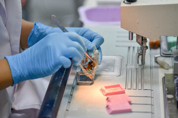
Hand in blue glove holding glass histology slides
Chronic gastritis of a human
Mildly inflamed colonic mucosa - microscopic image 20x
Ectocervical epithelium
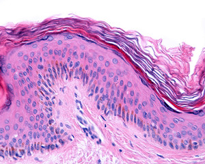
Thin skin. Hematoxylin-eosin
science microscope slide
Modern microscope and human tissue section slide with computer monitor show glandular image.Medical patholology and cytologytechnology concept.Selective focus.
Histology slide of human thyroid gland highlighting follicular cells colloidfilled follicles
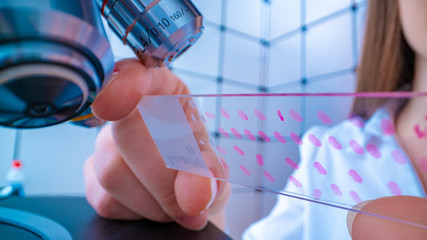
Young woman technician is examining a histological sample, a biopsy in the laboratory of cancer research
Education anatomy and Histological sample of Human under the microscope.
Colon biopsy showing a thickened collagen layer underneath the degenerating surface epithelium. This is a microscopic colitis called collagenous colitis. Microscopic view.
Hand in blue glove with histological rat liver, kidney an spleen tissue samples
rabbit epididymis cross section under the microscope showing spermatozoa inside its tubes - optical microscope x100 magnification
Fetal ovary: Oocyte development micrograph depicting leptotene and pachytene stages.
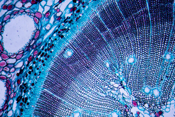
Microscopic image of Pine Stem (cross-section)
A pathologist is preparing a pathological biopsy for laboratory diagnosis of pathology.
Skin Tissue Layers: Epidermis, Dermis, and Hypodermis
Histology microscope image of Pacinian corpuscle of the dermis in skin (100x)
Archival Biopsy Complete Set
Cross section Human testis under microscope view.
Hand in blue glove holding glass histology slides
Skin papilloma of a human, highly detailed segment of panorama. Photomicrograph as seen under the microscope, 10x zoom.
Young female scientist preparing slides with paraffin-embedded sections for pathological analysis .
Close up photo of hand in white glove handing histological brain tissue sample
Thin skin. Hematoxylin-eosin
Mammalogist doctor examines woman breasts and lymph nodes during appointment. Skillful oncologist puncture of mammary glands of young patient under review ultrasound for diagnosis of breast cancer
Photomicrograph of the human scalp, showing histology of hair follicles, cut as a tangential cross section.
Scientist analyzing microscope slide at laboratory. Young woman technician is examining a histological sample, a biopsy in the laboratory of cancer research
Young woman technician is examining a histological sample, a biopsy in the laboratory of cancer research
Education anatomy and Histological sample of Human under the microscope.
Lung adenocarcinoma, light micrograph, photo under microscope
Hand in blue glove with histological liver tissue sample
microscopic section of Testis T.S tissue
Purkinje cells cerebellum, histology microscopic view neurons
Microscopic image of Pine Stem (cross-section)
rabbit ovary cross section under the microscope showing tunica albuginea, primordial follicles, primary follicles, secondary follicles, tertiary follicle and cortical stroma - optical microscope x100
Doctors or scientists are researching human organs tissue and blood samples, using microscope and reagents. A concept of histology service. Vector flat illustration, tiny person concept.
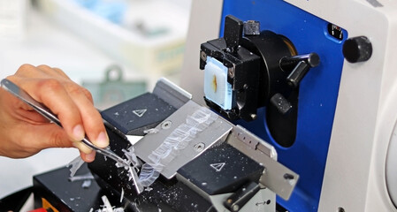
Scientific apparatus for cutting tissues and analyzing them in the microscope. Microtome.
Small glasses with pieces of histological biopsies on white background. The number 128364 can be read in the glasses. Small metal clamp.
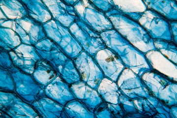
Onion cells at the microscope
Hand in blue glove holding glass histology slides
Histology of human tissue, show arteriolar hyaline degeneration of kidney as seen under the microscope, 10x zoom
Closeup of a microscope slide with stained tissue biopsy under the microscope. Cancer diagnosis concept. Medical concept.
Laboratory Set 04 Object Isometric
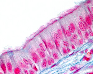
Respiratory epithelium
Microscopic photograph of a glomerulus in human kidney.
Colon cancer microscopic photography, magnification x40
Cerebellum cross section tissue under the light microscope.
Education Spinal cord  and Motor Neuron under the microscope in Lab.
Education Spinal cord and Motor Neuron under the microscope in Lab.
Acute myocardial infarction, histology of heart tissue, light micrograph. Area of infarct is paler than than the relatively viable area of heart muscle
Hand in blue glove with histological rat liver, kidney an spleen tissue samples
Paraffin Embedded Tissue Blocks of Cancer on iSolated White Background
microscope slide on a hand. Toned photo
Hand in blue glove with histological liver tissue sample
Scientist in blue gloves hand holding glasses slide breast tissue on out of focus cancer tissue picture background.Pathologist hand in glove holding glass organ samples.Histological examination.
Histology image of human fallopian tube showing ciliated simple columnar epithelia and mucous secreting cells (100x)
histology
histology
Microphotography of root of onion.
Modern laboratory interior out of focus
Hand in blue glove holding glass histology slides
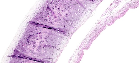
Histology of human tissue, show tracheitis as seen under the microscope
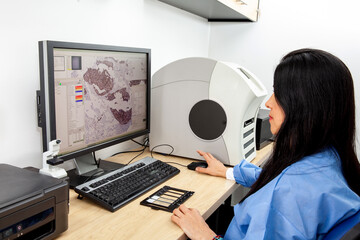
Young female scientist scanning microscope slides with tissue samples for pathology studies. Cancer diagnosis concept. Medical technology concept.
Normal duodenum mucosa showing long, slender villi. Inflammation is not increased. Microscopic view.
Melanocytes. Epidermis.
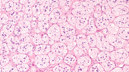
Microscopic image of an adrenal cortical adenoma, a benign tumor of the adrenal gland, which may produce hormones cortisol or aldosterone and lead to Cushing's syndrome or primary hyperaldosteronism.
Doctor shows information on blackboard: histology
Series of histology specimen slides
Xylem is a type of tissue in vascular plants that transports water and some nutrients.
Education anatomy and physiology of Tongue under the microscopic in laboratory.
Light micrograph of a liver showing cords made from hepatocytes and clear spaces between them called hepatic sinusoids and filled with red blood cells. Magnification 200x
Hand in blue glove with histological liver tissue sample
Cross section esophagus dog
Set of glass slides after thyroid fine needle aspiration biopsy, slides tissue biopsy for histology
Histology umbilical cord tissue
Scientist wear blue glove holding  human tissue block and out of focus modern microscope with digital camera and computer monitor show glandular image.Medical pathology and cytology technology
Scientist wear blue glove holding human tissue block and out of focus modern microscope with digital camera and computer monitor show glandular image.Medical pathology and cytology technology
histology of liver with labeled 3d Illustration
Visual Representation of Bone Marrow Microenvironment Exploring Cellular and Structural Components. Concept Bone Marrow Microenvironment, Cellular Components, Structural Components
Detailed pink agate texture background
Histological experiment
Hand in blue glove holding glass histology slides
Prostate Cancer(TURP): Microscopic image of prostatic tissue, adenocarcinoma, malignant neoplasm,atypical epithelial cells, malignant tumor
Closeup of a female scientist placing a sample on a transmission electron microscopy grid
Microscopic view of Mesothelioma cancer cells infiltrating the beige fibrous lung pleura.
Smooth muscle cells. Nuclei
Photomicrograph of lymphocytic colitis, a type of microscopic colitis, showing increased numbers of intraepithelial lymphocytes.  This condition is associated with watery diarrhea.
Photomicrograph of lymphocytic colitis, a type of microscopic colitis, showing increased numbers of intraepithelial lymphocytes. This condition is associated with watery diarrhea.
picture of histology human tissue with microscope from laboratory (not Illustration Designation)
Histological blade of cartilaginous tissue stained in Mallory forming a beautiful pattern with vivid colors.
Histological structure of pine stem vascular bundle under microscope.
Education anatomy and Histological sample Heart muscle Tissue under the microscope.
Lymph node metastasis, light micrograph of cancer that has spread to a lymph node
The head microscope on the background laboratory is a study human biology analyzes for non-compliance with the requirements organization public health at international level
Esophagus dog- Abstract background cell
abstract close-up macro image of a living cell, generative ai
Slide Cabinet for Storing Microscope Glass Slides
View in microscopic of normal human cervix cells.Squamous epithelial cells.Photo of Human skin cells.Superficial and intermediate epithelial cells.Cytology and pathology laboratory department.
Labium minus
Labium minus
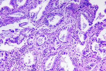
Well differentiated adenocarcinoma of a human


Why Wallsheaven is special?

30 Days on Returns

You can return or replace any product within 30 days without reason.

Free Express Delivery

Delivery is free for all of our products, wherever you are.

Eco Water-Based Inks

Our prints are odorless, ecological, and safe for children with vivid colors from natural dyes.

Simple Installation

Installing WallsHeaven's wallpaper mural is as easy as making paper airplane.

Artist Support

By choosing our photos, you will support the artists from all over the world.