
WallsHeaven makes decorative wall murals in high quality printing

24,790 Disease+nucleus+microscope Wall Murals - Canvas Prints - Stickers

Myocytes, Smooth muscle cell tissue
Human mammary gland cells under the microscope
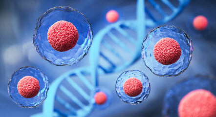
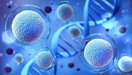
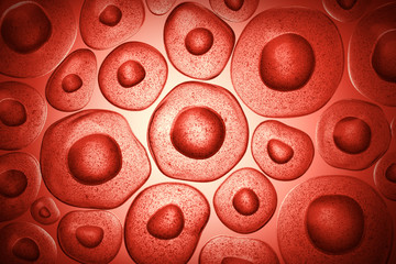
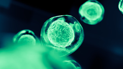
3d rendering of Human cell or Embryonic stem cell microscope background.
Red Cancer Cell Illustration
Synapse
Synapse
A microscope view of mitosis in cells. A background in biology
cells background
cells background
Parasitic protozoans Toxoplasma gondii
Cancer cells spreading
Background of human ovarian cyst cell tissue under microscope in pathology laboratory. View microscopically positive cervix adenocarcinoma, tissue sections by H and E
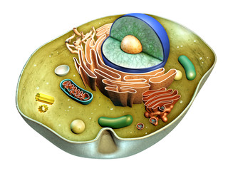
Cell structure
Cell structure
A detailed close-up of blue, microscopic bacteria floating in a dark environment.
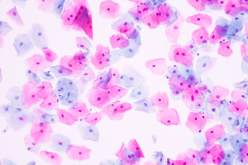
Normal squamous epithelial cells of cervical human view in microscopy.Cytology criteria from pap smear.Medical background concept.
Single red oval in liquid under microscope
This is a microscope used for the detection of pathogens such as bacteria, fungi, protozoa. and malaria etc. Suitable for scientists Medical technologist and medical scientists
 Viral Infection - A Close-Up Look at the Battle Within
Viral Infection - A Close-Up Look at the Battle Within
Microscopic bacteria. Science illustration. 3D render of infectious virus. Microbiology. Cells and dna. Very small molecules. Human health research. Abstract macro of medical. Microbe molecules.
blast cells in Leukemia
Education anatomy and Histological sample Heart muscle Tissue under the microscope.
Histology of human cardiac muscle under light microscope view for education.
Human mammary gland cells under the microscope
3d rendering of Human cell or Embryonic stem cell microscope background.
Abstract Spider Web Blood Vein Black And White Background
Targeted Cancer Therapy. Precision Attack on Malignant Cell
Cancerous tumor cells attacking the body at close range. Human biology microscope.
cells background
cells background
Chlamydia trachomatis bacteria, 3D illustration showing reticulate bodies of Chlamydia forming intracellular intracytoplasmic inclusions (small red) near the cell nucleus (purple)
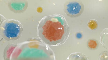
Human cells or Embryonic stem cells microscope background. The blue nucleus of the bacterium is surrounded by a transparent sphere. Medicine scientific concept. Generative AI
Confocal microscopy imaging of two cancer cells. Cytoskeletal proteins in red, nucleus in blue
Microscope red blood cells and virus Bacteria, infection 3d rendering, health care, medical and science concept. particle in blood, human body science biology
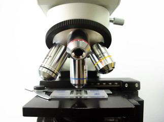
Close-up of a microscope
Backgrounds of human cells tissue of lung human under the microscope in pathology lab.View in microscopic of ductal cell carcinoma, adenonocarcinoma from cancer, tissue section by H and E stain.
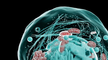
Schematic 3D depiction of a cell with a nucleus inside
This is a microscope used for the detection of pathogens such as bacteria, fungi, protozoa. and malaria etc. Suitable for scientists Medical technologist and medical scientists
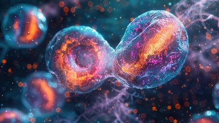
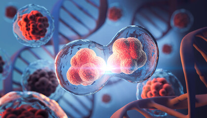
phase of cell division It shows that the cell divides to form 2 cells.
Confocal microscopy of fibroblast cells
blast cell on blood smear.
Cells with nucleus on scientific background
human cell
human cell
Cells through electron microscope (shallow dof)
3d rendering of Human cell or Embryonic stem cell microscope background.
Human acute leukemia cell line nuclei
Viral structure. Viral particle is made up of a nucleus of nucleic acid (DNA or RNA) surrounded by a protein coat. Conceptual illustrative virus.
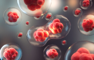
3d illustration of colorful cell division, cell membrane and splitting nucleus
Letter or word LABS GUMMY font with Microscope image graphic icon logo design abstract concept vector stock. Can be used as a symbol related to the laboratory.
Chlamydia trachomatis bacteria, 3D illustration, the causative agent of chlamydiosis
Histology of human cardiac muscle under light microscope view for education.
3d rendering of Human cell or Embryonic stem cell or vitamin microscope background. Cellular Therapy and Regeneration 3D illustration.
Cells in the microscope
Virus molecule microscope bacteria microbiology science study analyzing disease microorganism cell nucleus pathogen examining health pandemic, in dark style, black background
Cancer cell inside the body, Generative AI
Trichomonas in pap smear on white background view in microscopic.Medical background.Cytology and pathology laboratory department.Sexually transmitted diseases.Magnification 600 X.
bacteria or cells under the microscope. Multiplication of bacteria or infection. Microbes. Bacteria. attacking cells
This is a microscope used for the detection of pathogens such as bacteria, fungi, protozoa. and malaria etc. Suitable for scientists Medical technologist and medical scientists
The microscopic view of ovarian cancer cells, showing the nuclei in blue and the cytoplasm in pink, highlighting the s of abnormal cells that constitute malignant tumor.
Confocal microscopy of fibroblast cells
Mesothelial malignant cells with multiple nuclei
T Cells attacking Cancer Cells 3D illustration
Gills of fish under the microscope for Education.
Photomicrographic view of histological stained slide showing carcinoma. Adenocarcinoma
3d rendering of Human cell or Embryonic stem cell microscope background.
Real fluorescence microscopic view of human skin cells
3d rendering of Human cell or Embryonic stem cell microscope background
chromatin diagram labeled
Letter or word G in Microscope image graphic icon logo design abstract concept vector stock. Can be used as a symbol related to laboratory or initial
Chlamydia inclusion in human cells. 3D illustration showing group of chlamydial elementary bodies (red) near the nucleus (violet) of the cell
Histology of human cardiac muscle under microscope view for education.
Protozoa most common diseases set
Fluorescence Microscope image of Bovine Pulmonary Artery Endothelial Cells BPAE stained for mitochondria, phalloidin, and nuclei undergoing mitosis
Cell division during mitosis under a microscope
Colorful vivid bacteria and viruses microscope macro shot.
View in microscopic of koilocyte cell criteria of HPV (Human Papilloma virus) infection.Pap smear for woman.Medical background concept.
Red cells virus microscope from inside body with 3d rendering.
This is a microscope used for the detection of pathogens such as bacteria, fungi, protozoa. and malaria etc. Suitable for scientists Medical technologist and medical scientists
A patient has split skin transplanted on the abdomen to cover a wound
Fibroblast cells mebrane labeled with fluorescent dye
Abnormal neutrophil in pleural fluid smear.
T Cells attacking Cancer Cells 3D illustration
Light microscopic picture show erythrocyte, giant platelet, neutrophil, lymphocyte, nucleated red blood cell and polychromasia in the peripheral blood of cat with regenerative anemia.
Probiotic beneficial bacteria, normal intestinal microflora, microorganisms used as probiotics, yoghurts, healthy food, Lactobacillus, Bifidobacterium, 3d rendering
3d illustration of Embryonic stem cells under a microscope, Cellular therapy background.
Confocal microscopy of fibroblast cells
HeLa cervical cancer cells
The cell nucleus was believed to be elastic like a rubber ball. Generated with AI
A medical image of a leukemic blood smear, with an overabundance of immature white blood cells
cells seen at microscope
Squamous epithelial cells under microscope view for education histology.
Chlamydia inclusion in human cells. 3D illustration showing group of chlamydial elementary bodies (red) near the nucleus (violet) of the cell
Fluorescence Microscope image of Bovine Pulmonary Artery Endothelial Cells BPAE stained for mitochondria, phalloidin, and nuclei undergoing mitosis
A microscopic image of a human virus attacking a healthy immune system cell with colorful antibodies. Cellular therapy and regeneration. Research of stem cells, immunology and longevity.
View in microscopic of abnormal human cervix cells.Koilocyte cell criteria of Human Papilloma Virus (HPV) infection from pap smear slide.Cytology and pathology laboratory department.Magnification 400X
The tissue of a biological organism is affected by two types of cancer cells. Electron microscopy. Concept image of mutational processes under the influence of mutagens
Creative image of embryonic stem cells, cellular therapy, 3d illustration
A patient has split skin transplanted on the abdomen to cover a wound
cancer cells under a microscope. Tissues affected by cancer cells under the microscope, Science. Cancer drugs. chemistry


Why Wallsheaven is special?

30 Days on Returns

You can return or replace any product within 30 days without reason.

Free Express Delivery

Delivery is free for all of our products, wherever you are.

Eco Water-Based Inks

Our prints are odorless, ecological, and safe for children with vivid colors from natural dyes.

Simple Installation

Installing WallsHeaven's wallpaper mural is as easy as making paper airplane.

Artist Support

By choosing our photos, you will support the artists from all over the world.