WallsHeaven makes decorative wall murals in high quality printing

24,790 Disease+nucleus+microscope Wall Murals - Canvas Prints - Stickers

A vector representation of Multiple Myeloma cells, science, detailed -
Detailed 3D Render of a Vibrant Virus Particle with Blue Nucleus
A close-up of a bone marrow cell with a red bone marrow lining.
The image is a close up of a white surface with a blue wave pattern. The surface is illuminated with light, creating a sense of depth and movement. Scene is serene and calming
Photomicrograph of a soft tissue tumor in the dextra manus area in the anatomical pathology laboratory
Close-up of lung tissue under a microscope showing alveoli and capillary networks
Macro microscopic model of inflammation Neutrophil in blood 3D illustration
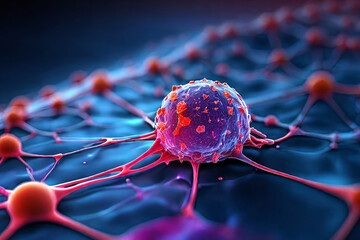
Cancer cell insight. Graphic representation captures the essence of medical science and healthcare. Ideal for websites, blogs, and print media. Explore the microscopic world.
A cluster of embryonic stem cells in 3D, with each cell glowing softly, revealing the nucleus and internal organelles under a microscope.
An immunofluorescence microscopy image depicting the distribution of Clara cells in bronchioles, showing their club-shaped morphology and secretion
Observing cancer cells under a microscope Scientific image intended for use in cancer medicine, chemistry, and biology.
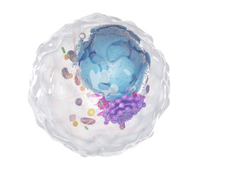
Human cell under a microscope. Embryonic stem cell components, nucleus and organelles, and plasma membrane. Medicine, microbiology, DNA, molecule.
blood through narrow passages, motion of blood cells
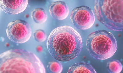
3d rendering of Human cell or Embryonic stem cell microscope background.
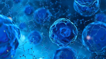
blue cell life biology medicine scientific molecular research background
Microscopic Close-Up of Toxoplasma Protozoan Parasite
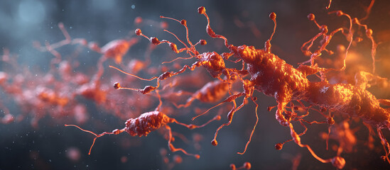
A microscopic view of a neuron with a glowing nucleus and dendrites.
Cross-section of the colon affected by colorectal cancer, showing tumors and their impact on colon function and health.
Microscopic View of Red Blood Cells in a Chain
Acanthamoeba amoeba and zoomed neutrophils
Niels Bohr's Atomic Model Bohr's model revolutionized our understanding of atomic structure and electron energy levels.
trichomonas microscopic view sexually transmitted disease STD
Genome wellness and fitness for transplant candidates, genome wellness fitness, high-tech genetic screening
 Human cell. Embryonic stem cell microscope. Components of Eukaryotic cell, nucleus and organelles and plasma membrane. Medicine, microbiology, dna, molecule. Generative AI
Human cell. Embryonic stem cell microscope. Components of Eukaryotic cell, nucleus and organelles and plasma membrane. Medicine, microbiology, dna, molecule. Generative AI
Human cervical cell tissue under a microscope in a pathology laboratory. Microscopic view of fibrosing mammary adenosis, hematoxylin eosin staining
A soft and calm scene with a light blue background, featuring very small molecular structures.
A vector representation of Non-Hodgkin's Lymphoma cells, science, detailed
Microscopic view of a virus showing detailed cellular structure in vibrant colors
A microscopic view of red blood cells and their structures
Embryonic stem cells colony under a microscope. Cellular therapy and research of regeneration and disease treatment in seamless 3D illustration. Biology and medicine of human body concept. 4K
Observing cancer cells under a microscope Scientific image intended for use in cancer medicine, chemistry, and biology.
A schematic 3D representation of an inside of a cell containing a nucleus
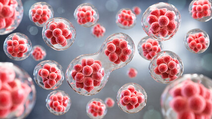
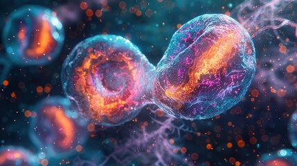
D Illustration of a Cell Dividing with Multiple Nuclei
Electric neurons firing through a vibrant abstract space of light
Abstract microscopic virus or bacteria. Microbiology and biology. 3D render. Scientific background
Light microscope Pigmentary of liver pathology.
Tissue sample of Bone human, Elastic cartilage human and Red bone marrow under the microscope in Lab.
A microscope view of a ebola virus cell
Microscopic Exploration A 3D Render of a Microscope and Bacteria, science, microscope, bacteria, research
A detailed 3D illustration of a cancer cell nucleus, highlighting its structure and complex processes. The image symbolizes cellular division, disease progression, and the fight against cancer.
cells, bacteria and microorganisms magnified under a microscope
A vivid depiction of cellular structures floating within a fluid environment, featuring vibrant colors and intricate textures, evoking a sense of microscopic life.
3d rendered illustration of a healthy human cell
 Human cell. Embryonic stem cell microscope. Components of Eukaryotic cell, nucleus and organelles and plasma membrane. Medicine, microbiology, dna, molecule. Generative AI
Human cell. Embryonic stem cell microscope. Components of Eukaryotic cell, nucleus and organelles and plasma membrane. Medicine, microbiology, dna, molecule. Generative AI
Human cervical cell tissue under a microscope in a pathology laboratory. Microscopic view of fibrosing mammary adenosis, hematoxylin eosin staining
A pill is surrounded by a cloud of red and orange particles
A microscopic view of multiple cells with a prominent nucleus and other organelles, surrounded by a fluid. The cells are in a light blue background.
A 3D rendering of a human cell or embryonic stem cell microscope background.
blast cells in Leukemia
A microscopic view of green algae. AI.
Observing cancer cells under a microscope Scientific image intended for use in cancer medicine, chemistry, and biology.
Embryonic stem cells colony under a microscope. Cellular therapy and research of regeneration and disease treatment presented as seamless 3D illustrations. 4K biomedical image.
Human cell structure: nucleus, cytoplasm, golgi, organelles mitochondria, ribosomes, lysosomes, cell membrane. Genetic DNA replication, RNA, protein synthesis, biology medical science. 3D cell anatomy
A blue and white image of a cell with a blue nucleus and a green cytoplasm
Intricate microscopic view of cellular structures showcasing vibrant orange nuclei within a web of delicate fibers.
Macro of infected virus cells, dividing the body moving inside the body, visible through a microscope
Tissue sample of Bone human, Elastic cartilage human and Red bone marrow under the microscope in Lab.
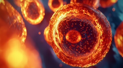
A red and orange blob of something that looks like a cell
microscopic view of a neuron, revealing the complex network of dendrites and axons that allow for communication between cells
Blue glowing cells fractal
 endless possibilities and complexity of the miniature world of cells
endless possibilities and complexity of the miniature world of cells
Close up influenza virus in blood vessel. Red abstract plexus wireframe Coronavirus background. Science and medical concept. Micro nucleus of Corona virus cell in human body. 3D illustration graphic
Close-up of colorful, abstract shapes representing cells under a microscope
Fluorescent Imaging immunofluorescence of cancer cells growing in 2D with nuclei in blue, cytoplasm in red and DNA damage foci in green
3d rendering of Human cell or Embryonic stem cell microscope background.
This is a microscope used for the detection of pathogens such as bacteria, fungi, protozoa. and malaria etc. Suitable for scientists Medical technologist and medical scientists
3d rendering of Human cell or Embryonic stem cell microscope background.
Human cell or Embryonic stem cell microscope background.
Targeted Cancer Therapy. Precision Attack on Malignant Cell
Detailed 3D rendering of a mitochondrion, the powerhouse of the cell showing internal structures like cristae in vibrant blue color.
The tissue of a biological organism is affected by two types of cancer cells. Electron microscopy. Concept image of mutational processes under the influence of mutagens
An enlarged view of a cancerous tumor. A macro view of a cancerous tumor.
 Cancer cells observed under a microscope
Cancer cells observed under a microscope
A blue and white image of a cell with a blue nucleus and a green cytoplasm
a microscope with a cell phone on it, Showcase the technology behind a fluorescence microscope
A vector representation of Myelodysplastic Syndrome cells, science, detailed
A colorful, abstract image of a brain with many holes and a blue background
Visual representation of cell apoptosis, cell breaking down with colorful fragments scattering, cell apoptosis, biology, pathology
cells seen at microscope
Cartilage cells under the microscope
living worlds under the microscope, where cells, bacteria and microorganisms
Viral structure. Viral particle is made up of a nucleus of nucleic acid (DNA or RNA) surrounded by a protein coat. Conceptual illustrative virus.
Microscopic Purple Cellular Structures
Creative image of embryonic stem cells, cellular therapy. 3d illustrations. Generative AI
400x magnification of Coleastrum, Scenedesmus, Euglena, and another green algae with selective focus
3d rendering of Human cell or Embryonic stem cell microscope background.
Cells through electron microscope (shallow dof)
3d rendering of Human cell or Embryonic stem cell microscope background, Geneative AI
 A closeup view of a neuron with a glowing nucleus illustrating the complexity of the nervous system
A closeup view of a neuron with a glowing nucleus illustrating the complexity of the nervous system
Education anatomy and Histological sample Heart muscle Tissue under the microscope.
A microscope used to view cells, embryonic stem cells, and cellular therapies.
3D rendering interpretation of virus on cells, The nucleus and cytoplasm.
3D Illustration of a DNA filled liquid being injected into an egg cell nucleus
Green bacteria with nucleus and cilia multiply, ideal for health and biology concepts. Explore microorganisms in this educational graphic
Sizes of cells drawn on a logarithmic scale, indicating the range of readily resolvable objects in the light and electron microscope.
Detailed image of a single-celled organism like a paramecium under a microscope highlighting its cilia and nucleus
phase of cell division It shows that the cell divides to form 2 cells.


Why Wallsheaven is special?

30 Days on Returns

You can return or replace any product within 30 days without reason.

Free Express Delivery

Delivery is free for all of our products, wherever you are.

Eco Water-Based Inks

Our prints are odorless, ecological, and safe for children with vivid colors from natural dyes.

Simple Installation

Installing WallsHeaven's wallpaper mural is as easy as making paper airplane.

Artist Support

By choosing our photos, you will support the artists from all over the world.