



WallsHeaven makes decorative wall murals in high quality printing

24,790 Disease+nucleus+microscope Wall Murals - Canvas Prints - Stickers

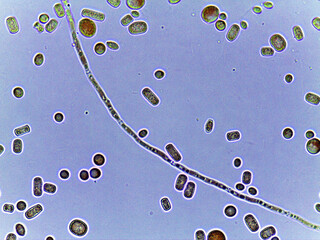
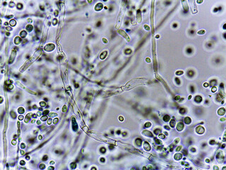
aspergillus fumigatus under the microscope. a species of higher mold fungi of the genus aspergillus,
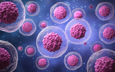
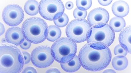
a group of cells, each with a nucleus and cytoplasm.
neuron cells nervous system closeup, abstract blue glitter neuron cells in brain artistic background
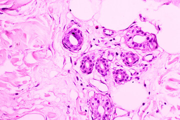
backgrounds of human cells tissue of cervix under the microscope in pathology lab.view in microscopi
this image encapsulates a surreal representation of cells glowing in various hues, invoking a sense
digital visualization of a neuron that transmits nerve impulses. neuron cells with glowing link knot
photo of fungi spores and mycelium under the microscope
backgrounds of human cells tissue of cervix under the microscope in pathology lab.view in microscopi
abstract microscopic virus or bacteria. microbiology and biology. 3d render. scientific background
a cluster of dna strands are shown in a blue and purple color. the strands are connected to each oth
cell mutation, highlighting the differences between normal and mutated cells in a vibrant cellular l
colorful microscopic bacteria and viruses floating in a vibrant environment, showcasing various shap
microbiology macrophotography
a closeup view of a single young erythrocyte with a large nucleus taking on a characteristic biconca
a colorful image of a cluster of red and blue cells
a microscopic illustration of the steps involved in somatic cell nuclear transfer from the isolation
probiotics substance. microbiota background. beneficial bacteria in body. blue medical background. p
a collection of white spheres in a blurry image
stem cell research advances our understanding of cellular therapy and regeneration, delving into mic
purple cell biology clipart, medical illustration, png, transparent background, cut out background,
a microscopic view of human cells or neurons
two glowing red neurons in a blue background
a vector representation of acute lymphoblastic leukemia cells, science, detailed
a vector representation of multiple myeloma cells, science, detailed -
a vector representation of myelodysplastic syndrome cells, science, detailed
a close-up view of an abstract molecular structure, illuminated with bright blue and orange lights.
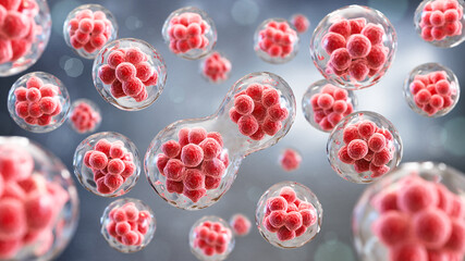
microscopic view of yellow cells dividing human or embryonic stem cells. concept biology, cell divis
3d rendering of human cell or embryonic stem cell microscope background.
3d of the fertilization of the embryo in the uterus
plant cell under a microscope, showing genetic edits and disease effects in vivid detail, no humans,
human skin cells under a microscope
microscopic close-up of toxoplasma protozoan parasite
a man's body is covered in green vines and leaves
an illustration of bacterial cell division, explaining microbial proliferation
paps smear: nilm with reactive cellular changes associated with inflammation.
3d probiotic medical background, microbiology microbiota bacillus blue banner microflora render. hea
scientist using advanced microscope in modern laboratory for research and analysis. high-tech lab eq
a close up of a pinkish red blob with a lot of bubbles
a close up of a red cell with a black center
3d rendering of human cell or embryonic stem cell microscope background.
zellen in langgezogener form unter dem mikroskop, bunte farben, vor schwarzem hintergrund, querforma
creative image of embryonic stem cells, cellular therapy. 3d illustration, generative ai
the image is a close up of a blue and white pattern with many small circles
fluorescent cells. experiment in laboratory clinic showing growth of aggressive cancer cells
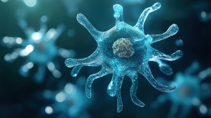
abstract biological cell structure with tentacle like extensions on golden background
close-up of bone tissue under a microscope showing the detailed cellular structure and osteocytes
a close-up photo of an amoeba under a microscope, highlighting its nucleus and pseudopods
detailed microscope view of a single-celled creature, such as a paramecium, displaying its nucleus a
a detailed image of a somatic cell nucleus being transferred into an enucleated oocyte for the purpo
a microscopic snapshot of the somatic cell nuclear transfer technique in action with a somatic cell
a microscopic illustration of the steps involved in somatic cell nuclear transfer from the isolation
a close up of a cell with a red dot in the middle
microscopic view of immune cells, actively targeting and engulfing pathogens.
microscopic view of malignant b cells with irregular nuclei and abundant cytoplasm in a lymph node b
pathogen cell, bacteria and virus isolated on transparent background
yellow fat, like obesity cells, under the microscope
illustration of cancer cells under a microscope
a closeup view of a single young erythrocyte with a large nucleus taking on a characteristic biconca
a close up of many small yellowish brown circles
a colorful cluster of cells with a blue center
microscopic view of red blood cells and cellular structures
vector illustration of cll cell biology
a close up of a greenish color with many small bubbles
micrograph of a virus attacking a healthy immune system cell. stem cells, immunology, and longevity
neural impulses and connections in human nervous system
epstein-barr dna herpes virus infecting a b lymphocyte, electron micrograph. ebv, herpesvirus 4 cell
visual representation of cell apoptosis, cell breaking down with colorful fragments scattering, cell
observing cancer cells under a microscope scientific image intended for use in cancer medicine, chem
microscopic bacteria in blue
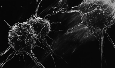
microscopic view of cancer cells with glowing nuclei and intricate network of fibers in a dark envir
trigger point in a muscle fiber, highlighting its striated appearance
close-up of reproductive cells under a biological microscope, showing intricate details in soft blue
a 3d illustration of a red cell with a glowing nucleus and a strand of dna.
3d rendering of human cell or embryonic stem cell microscope background.
eosinophils: a close-up of eosinophils, characterized by their bilobed nuclei and large granules sta
tissue sample of bone human, elastic cartilage human and red bone marrow under the microscope in lab
microscopy: confocal imaging of fibroblast cells in red, green and blue colors
microscopy: confocal imaging of fibroblast cells in red, green, and blue
coronavirus covid 19 microscopic view showing corona virus spikes attached to cell nucleus
human cervical cell tissue under a microscope in a pathology laboratory. microscopic view of fibrosi
a microscope view of a ebola virus cell
3d rendering of human cell or embryonic stem cell microscope background.
microscopic view of cells and viruses
a detailed view of a cancer cell nucleus undergoing division, showcasing the complex process of cell
an electron microscope image revealing the abnormal morphology of cancer cells, with distorted nucle
cell division. cellular therapy. 3d illustration on a dark background with images of cells under a m
a microscope of a cell, embryonic stem cells, cellular therapy and regeneration.


Why Wallsheaven is special?

30 Days on Returns

You can return or replace any product within 30 days without reason.

Free Express Delivery

Delivery is free for all of our products, wherever you are.

Eco Water-Based Inks

Our prints are odorless, ecological, and safe for children with vivid colors from natural dyes.

Simple Installation

Installing WallsHeaven's wallpaper mural is as easy as making paper airplane.

Artist Support

By choosing our photos, you will support the artists from all over the world.